Last week, our lab participated in the 23rd European Conference on Eye Movements, which took place in Ulm, Germany.
Our lab had a total of five presentations at the conference, three talks and two posters.
All presentations focused on the neural control of eye movements, which was an important addition to the conference’s other topics, like applied eye tracking and clinical biomarkers.
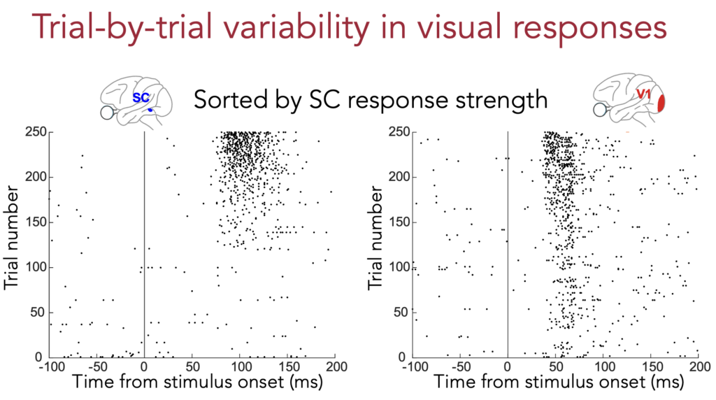
First off, Yue kicked off our talks in a symposium focused on neural mechanisms. She spoke about her simultaneous recordings of the superior colliculus (SC) and primary visual cortex (V1) of the same animals. In her prior work, she had shown how visual responses in the SC are much better related to eye movement timing than visual responses in V1 (like here and here). However, this raises a fundamental unanswered question because we know that V1 sends direct anatomical projections to the SC. So, does that mean that SC and V1 visual responses do not covary with each other across trials? Yue tackled this problem by simultaneously recording SC and V1 visual responses during simple fixation. She found that there was still some covariation between neurons in the two areas, consistent with the anatomy. However, the covariation was weak. More importantly, the covariation was strongest between deep SC neurons and V1, inconsistent with the anatomical projection (which favors more superficial SC neurons). This means that the covariation that Yue observed between the two brain areas is not due to anatomical drive from V1 to the SC. Instead, it suggests that the covariation likely reflects global brain-state changes that affect multiple brain areas simultaneously. This observation not only reconciles the question of anatomical connectivity, but it is also consistent with our earlier work on the links between either the SC or V1 and eye movement timing.
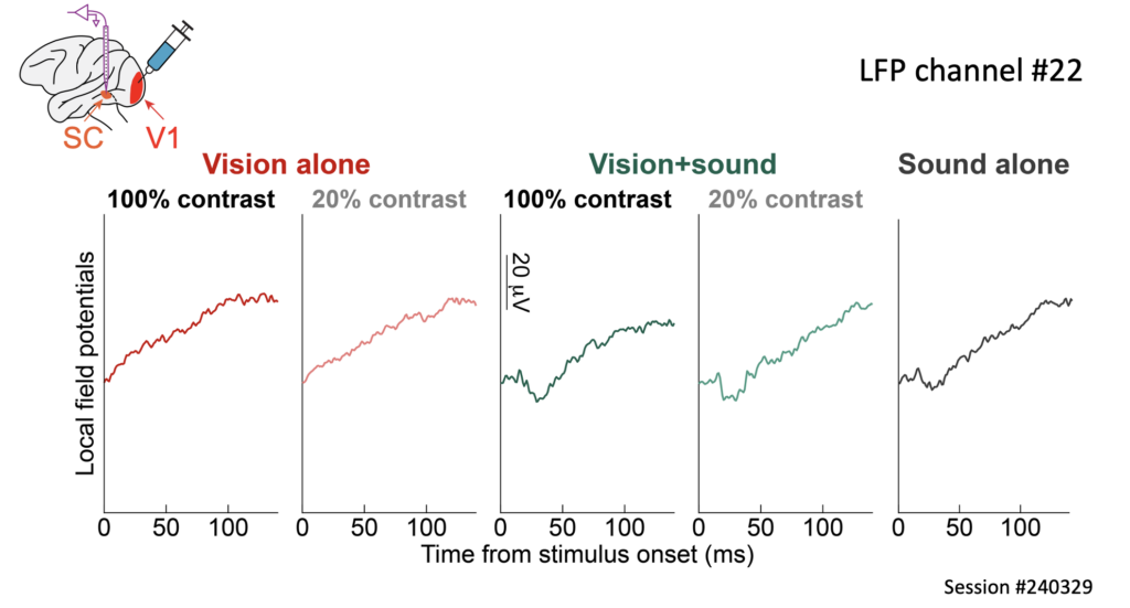
Next came Tanya’s talk. Here, she directly investigated the functional implications of a direct anatomical connection from V1 to the SC. She inactivated V1 and recorded from the SC (and also the neighboring inferior colliculus, IC). Remarkably, she found that there was still a weak visual signal in both the SC and IC that persisted despite the loss of V1 activity. Moreover, this weak signal was amplified with multisensory stimulation, and it even unmasked behavioral eye movement effects that were lost with visual-only sensory stimulation. Thus, even though the V1 pathway is dominant in the primate visual system, this does not deny that there are other pathways, such as direct retino-tectal projections, that provide very short-latency visual signals into the midbrain. These signals likely help in boosting behavioral responses to more ecologically relevant multisensory stimuli. More information about this work can be obtained from two very important papers by Tanya: here and here.
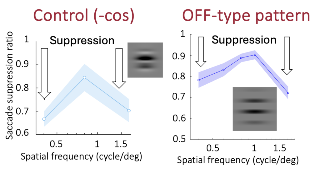
The third talk in the symposium was given by Wenbin. This time, he focused on how visual sensitivity in both the SC and V1 can be impaired around the time of saccadic eye movement generation. He used his recent discovery that dark contrasts are immune to saccadic suppression in V1 (still unpublished) to ask whether this asymmetry of V1 saccadic suppression can explain perceptual phenomena with grating stimuli (that have both dark and bright subcomponents). Such grating stimuli have been a work horse of vision science for many decades, because they allow controlling the spectral content of images in experiments. Wenbin also added a computational model of the early visual system to explain his results. This work is important because it continues our lab’s tradition of trying to understand the visual origins of saccadic suppression (such as in this paper)…..the surprises keep coming!
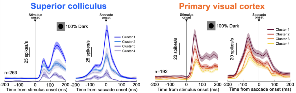
Shweta gave our lab’s fourth presentation at the conference, this time in the form of a poster. She continued her work on trying to link SC and V1 action potential waveform shapes to the functional properties of the neurons being recorded. She clustered waveforms purely based on their shape. Then, she explored the functional properties of the neurons in the different clusters. Remarkably, she found large differences in the discharge properties of the neurons in the different clusters, suggesting that the biophysics of individual neurons (which dictate action potential waveform shapes) also map onto distinct functional classes. For example, motor burst strengths in the SC were drastically different in the different clusters of SC neurons, within the same task and same animals. Similarly, visual response strengths in V1 were also drastically different across clusters. Importantly, her work shows that action potential waveform shapes are not the same throughout the whole brain, as might be implied from the existing literature. Rather, each area can have its own distinct neural discharge properties of the action potentials. For example, we found that SC waveforms were generally much thinner, on average, than V1 waveforms. This work is important because it, like Tanya’s work above, helps to bridge structural considerations (including anatomy and biophysics) to functional observations of brain function.
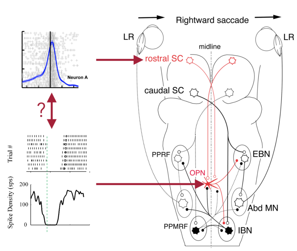
Finally, Ziad gave a poster on the motor properties of omnipause neurons (OPN’s) in the brainstem. This was the most motor poster of all of our presentations, and it addressed a large controversy that has persisted in the field of oculomotor control. Namely, it was suggested for more than 30 years that an excitatory projection from the rostral end of the SC to OPN’s is there to maintain these neurons’ tonic discharge during gaze fixation. However, this prevalent model cannot reconcile with the fact that the rostral SC emits strong motor bursts at the time of microsaccades, during which OPN’s should still pause instead of burst! Together with Antimo Buonocore and Maria, Ziad showed that the excitatory connection from the rostral SC to OPN’s is not there to prevent saccades; rather, it is likely used very strategically by the brainstem network, in order to achieve fine-scale oculomotor control of small and slow eye movements.