
We have a new paper just published in iScience!
In this paper, we investigated a fundamental question on why a brain structure like the superior colliculus (SC) should exhibit short-latency visual responses that are qualitatively almost identical to visual responses in the primary visual cortex (V1).
It is well known that the SC receives a direct anatomical connection from V1, but does that mean that SC visual responses are merely inherited from V1? Or, is the “image” formed in the SC at the time of visual stimulus onset computationally reformatted for another purpose than the “image” that is formed in V1 by the same stimulus?
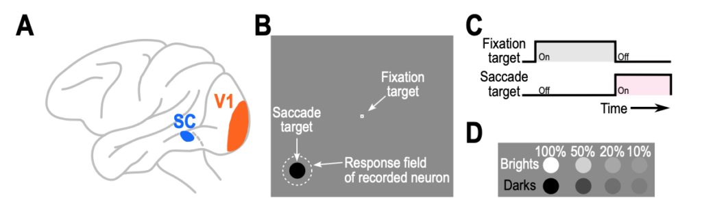
We explored this question by linking visual responses in both brain areas to rapid foveating eye movements that we employ to look at the world around us. Whenever a visual stimulus suddenly appears in the environment, an automatic reflex is usually to directly look at it and bring its image into the foveal region of the retina. Before such a foveating eye movement is triggered, both SC and V1 neurons would exhibit visual responses to the appearing eccentric stimulus, and we asked how the properties of these visual responses correlate with the so-called “saccadic reaction time”. By performing experiments in the very same animals, and with the very same stimuli, but in the two different brain areas, we could directly compare the relative differences between the two areas – without worrying about other confounds like differences in individual subjects, stimuli, or laboratory conditions. Such confounds could not be resolved based on the evidence from the literature so far, and that is exactly why our study was important to conduct.
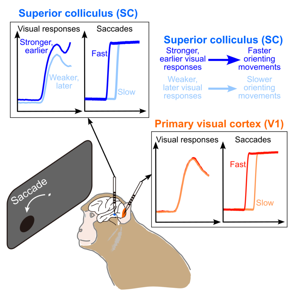
For a variety of different image contrasts and appearances, we found something remarkable: predicting saccadic eye movement reaction time from visual responses on any given trial is much better in the SC than in V1. Thus, SC visual responses are reformatted in a way that helps dictate, at least in part, how quickly a foveating eye movement might be triggered. This is consistent with our earlier hypothesis that the SC might possess what we called “a vision for orienting“, which is not necessarily the case for V1.
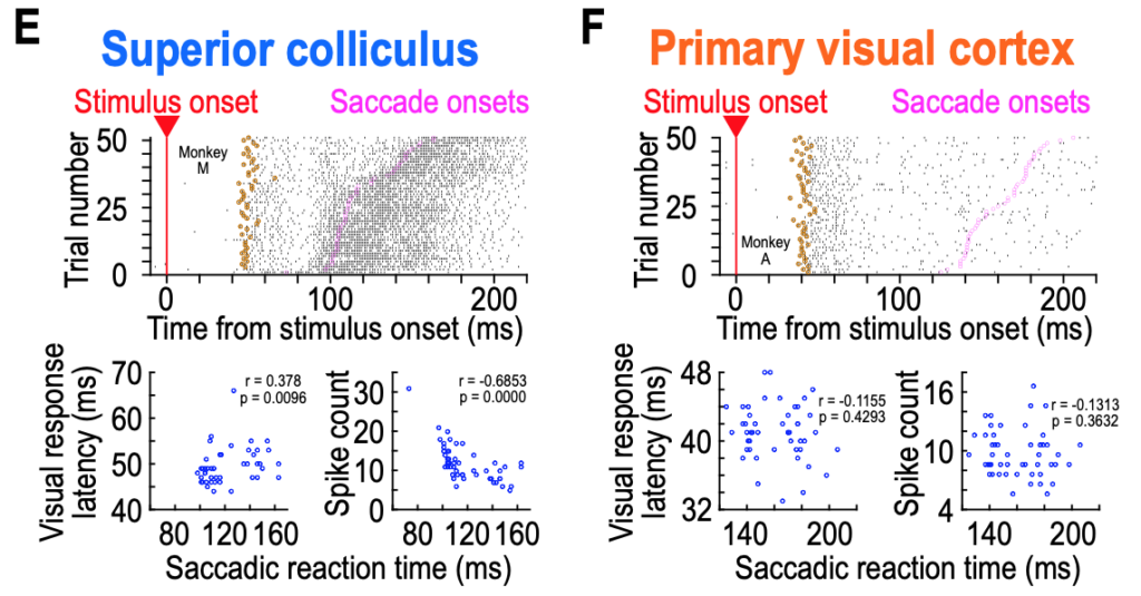
Across the entire population of SC and V1 neurons that we studied, this observation meant that visual responses to the very same stimulus in the SC could be very different on trials with either fast or slow eye movement reaction times, whereas V1 visual responses were not dramatically altered by when the subsequent saccade was triggered.
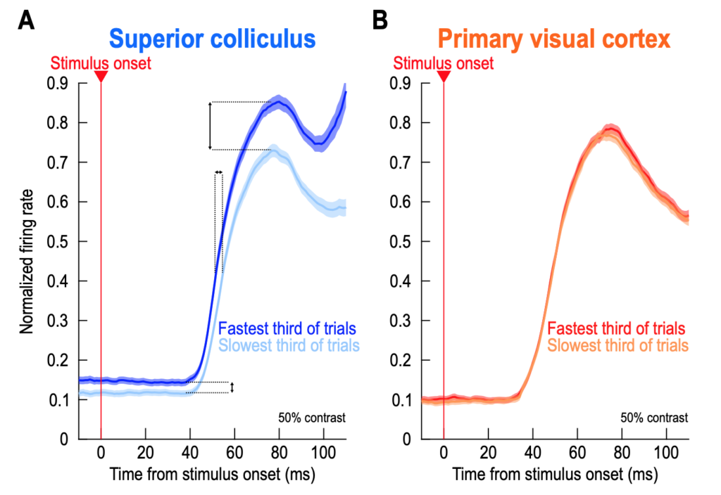
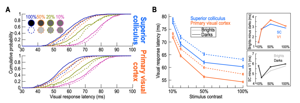
Finally, our paper was the first in the literature to document the average response latencies of V1 and SC neurons in the very same animals and with the very same stimuli. We found that SC visual responses were occurring systematically 5-6 ms later than V1 visual responses (consistent with the direction of anatomical connectivity between V1 and SC), but that differences within an area with different image properties were quantitatively more similar to each other (i.e. the image-dependent differences in visual response latencies were the same regardless of which brain area we were recording from).
This paper sets the stage for several important follow up studies that will continue our investigations of the similarities and differences between the SC and V1 in active vision!