We have a new very exciting paper at Nature Communications. The paper addresses a topic that we, like many others in the field of active vision, had been interested in for a long time, and it contains many exciting and unexpected discoveries in it. Indeed, it represents a real tour-de-force by graduate students Matthias Baumann and Saad Idrees.
In this paper, we explored the phenomenon of perceptual saccadic suppression. That is, whenever a brief flash is presented to the visual system around the time of a saccadic eye movement, perception of this flash is severely impaired. There have been many debates about how such suppression takes place. A prevalent theory was that the brain actively, through knowledge of its internal eye movement command, suppresses visual sensitivity. However, we hypothesized that this phenomenon could fundamentally be visual in nature without a need to know about the internally generated movement command.
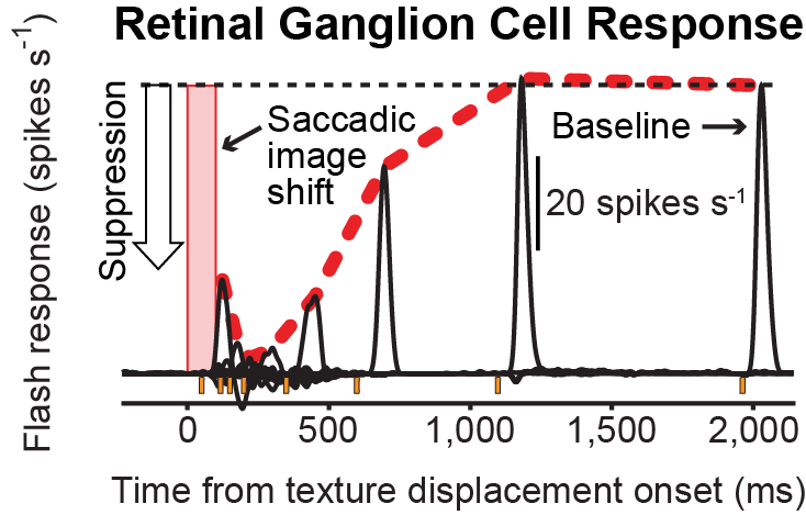
To test this, we isolated ex vivo retinal tissue from multiple animal species. We then “simulated” what a saccadic eye movement looks like from the perspective of the retina. A saccade is a rapid eyeball rotation. This means that the retina experiences a rapid global visual image flow every time a saccade is executed. We therefore translated images rapidly to the ex vivo retinal tissue, and we saw how retinal ganglion cells (the output neurons of the retina) then responded to brief visual flashes identical to those used in perceptual experiments. We found that retinal sensitivity was severely suppressed, and in a manner similar to previous reports of “saccadic suppression” in the literature.
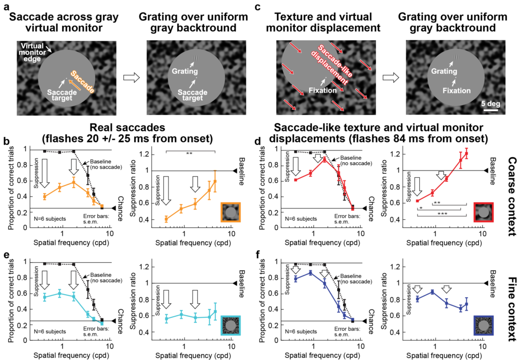
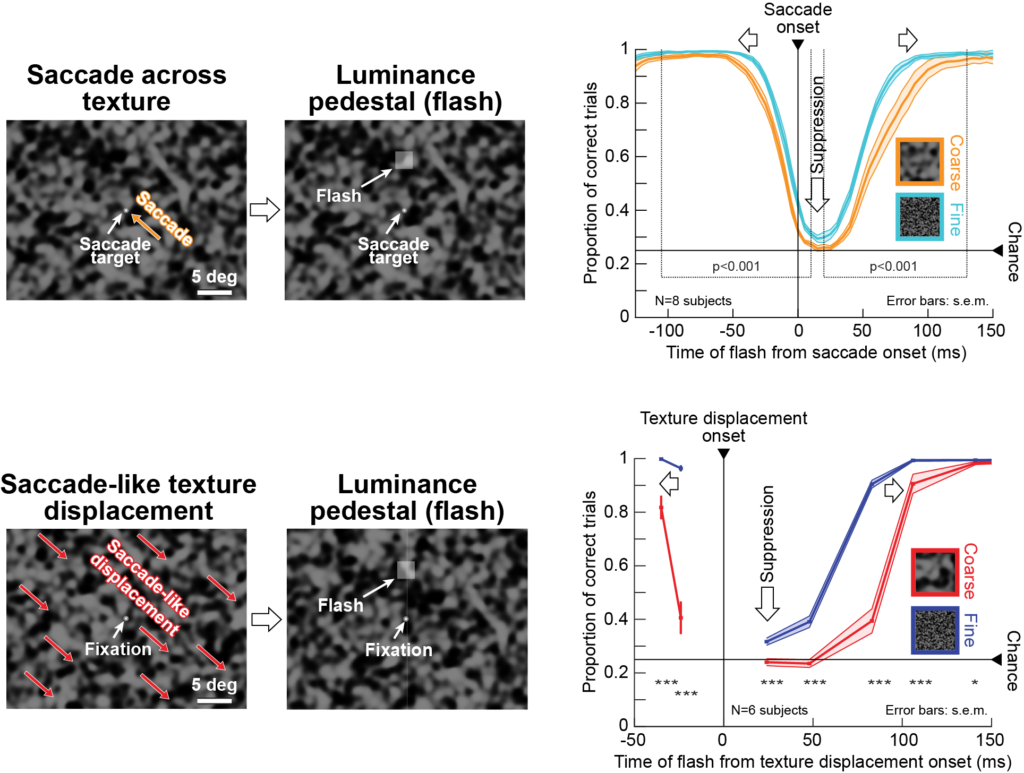
From these experiments, we also noticed something even more intriguing. If we modified the properties of the image that the retinal tissue was “seeing”, we could modify the properties of neural suppression. This gave us a strong prediction on how perceptual saccadic suppression should behave in human participants. We therefore tested the perception of these human participants under different image conditions. We found that, indeed, saccadic suppression depends on image statistics. More importantly, the dependence was identical whether the participants made real saccades (top row in the image below) or whether their retinae only experienced “simulated saccades” like the ex vivo retinal tissue in the experiments above (bottom row in the image below). And, the dependence was exactly like that we observed in the ex vivo animal retinal tissue. Therefore, with or without real saccades, “saccadic suppression” could still occur, as an outcome of general image processing operations performed at the very first stage of the entire visual system (the retina).
Armed with this insight, we finally made a daring hypothesis. In a classic study from 1995, which has often been used as a poster child for hypotheses of movement-related control of saccadic suppression, it was shown that when the brief flashes are now textured gratings of certain spatial frequencies, then only the lowest spatial frequencies are suppressed. This has been interpreted in the literature as movement-related commands being used to target specific types of cells in the early visual system for suppression. However, we used the same approach above of real versus “simulated” saccades in our human participants, and we found not only that this phenomenon can happen without any saccades at all, but that the dependence of saccadic suppression on low spatial frequencies can be so easily violated with very simple “visual” tricks in the image. Therefore, with or without a “movement command”, selectivity of saccadic suppression to low spatial frequencies (a very classic result in the field) may or may not happen. This means that even this classic study is describing a fundamentally visual phenomenon!
These results open many new interesting questions that we are now actively exploring. The full paper can be read here.