
We have a new paper now published in the Journal of Neuroscience. In it, we explored a phenomenon that we recently documented about ocular position drifts that occur during maintained gaze fixation.
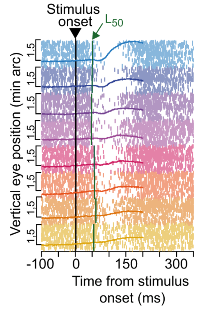
Ocular position drifts are generally the smallest known eye position displacements that cause retinal image shifts. These eye movements displace images on the retina by about the size of a single foveal photoreceptor, and we recently found that they are reflexively modulated by visual stimulus onsets (with very short latency). In the current study, we extensively characterized the properties of this stimulus-driven ocular position drift phenomenon.
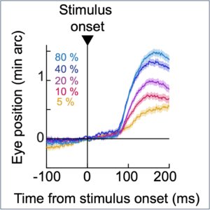
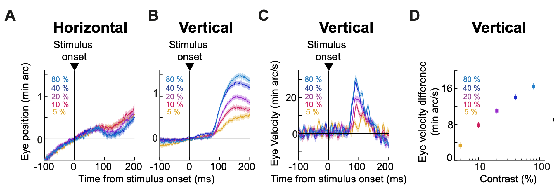
Like in our recent study of microsaccadic inhibition, we presented a series of image feature dimensions during gaze fixation, and we investigated how the ocular position drift response was altered. For example, we studied how the drift response depends on stimulus contrast. We found that its amplitude monotonically scaled with contrast. Similarly, stimulus size clearly modulated the drift response, with bigger stimuli generally causing larger and faster drift responses.
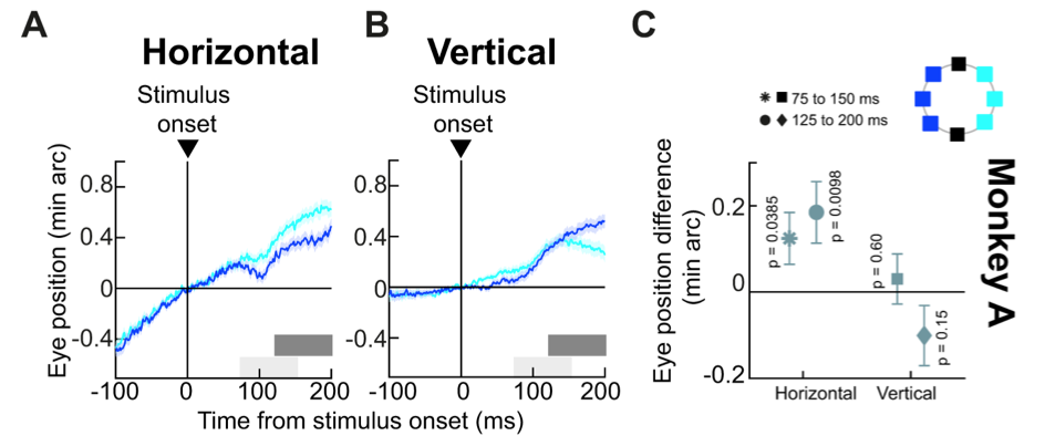
We performed extensive additional experiments, perhaps the most revealing of which was the one in which we presented localized peripheral flashes in the environment. We know since two decades of work on microsaccades that peripheral flashes modulate the time and direction of these small, but rapid eye movements. Remarkably, we now know that this also happens for the much smaller and slower ocular position drifts! This is a remarkable similarity with the other small eye movements that occur when maintaining gaze fixation: microsaccades. And, it also likely has implications, which we discussed a decade and a half ago, on the neurophysiological mechanisms driving ocular position drifts.
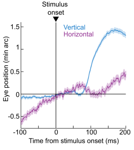
Because the drift response is also almost always accompanied by a strong upward component, we had previously hypothesized that it might be driven by the preference of the superior colliculus (SC) for the upper visual field. However, when we now tested this explicitly, we found that the upward component of the drift response still persists even if we exclusively presented stimuli only in the lower retinotopic visual field. This suggests that mechanisms other than SC stimulus-driven activity likely drive the upward component of the drift response. We are now exploring other potential brainstem mechanisms that might be involved.
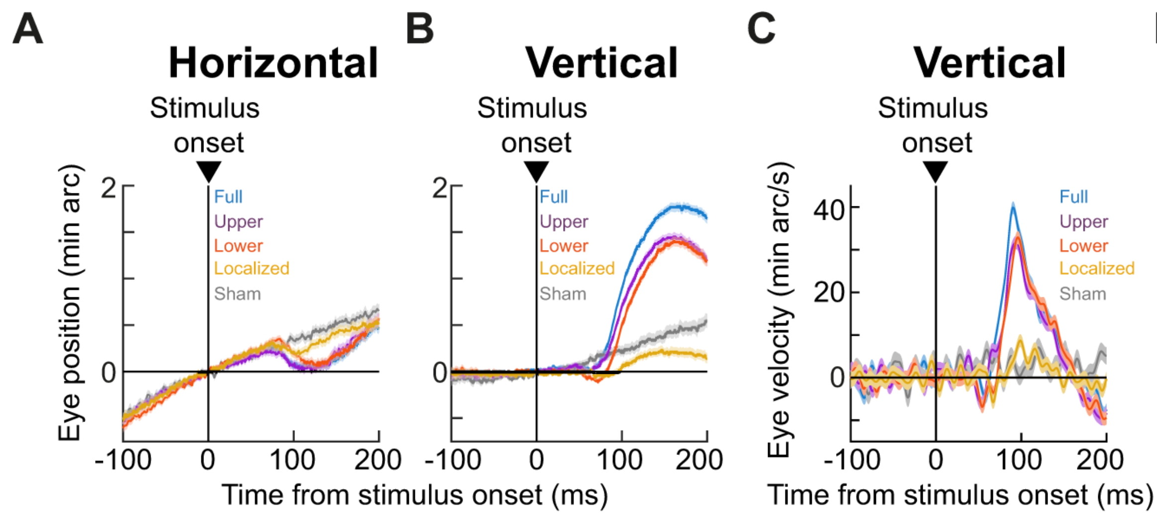
Finally, we related the drift response to the phenomenon of saccadic inhibition, which also happens inevitably with short latencies after stimulus onsets. We found a very strong temporal synchrony between saccadic inhibition and ocular position drift responses, suggesting that the two phenomena may be linked by the same underlying neuronal mechanisms. This helps inform some hypotheses that we have, which we are now actively testing in the lab.
Our massive paper has many more details, and it can now be read here.