
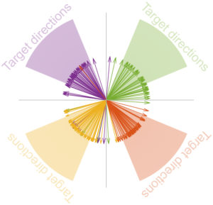
 We have a new paper in press at Nature Communications. In this paper, we tackled the issue of individual microsaccade generation. Microsaccades are overwhelmingly described in the literature, as well as in the public domain, as being involuntary, reflexive, and spontaneous eye movements. However, this contradicts a lot of evidence, including our very own prior research, showing how microsaccades can be related in very specific ways to cognitive state (such as attention). We, therefore, created a behavioral task called “memory-guided microsaccades” to demonstrate that any individual microsaccade can be generated at will, based on an abstract instruction, and without any visual guidance. The task involved subjects remembering a specific location within foveal visual space (i.e. very close to where the subjects were actually looking) and then waiting until an abstract instruction to “look” at the remembered location. We found that the subjects could easily generate these memory-guided microsaccades. Moreover, the movements were very spatially accurate. For example, this movie of eye position demonstrates an example trial’s result in slow motion, in which it is clear that the subject could easily generate a tiny, but spatially accurate, microsaccade to a remembered “blank” display location.
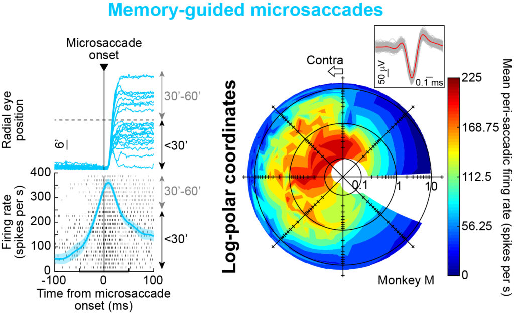 We then recorded neural activity from the superior colliculus (SC), a midbrain structure thought to be important for microsaccade generation. We found that the SC was still activated whenever a memory-guided microsaccade to a blank display location was generated. This means that microsaccades are not only voluntary, but they are supported by SC circuitry in the brain.
Another important advantage of studying memory-guided microsaccades was that we now had a behavioral task that very nicely dissociated the actual eye movements (microsaccades) from the visual stimuli guiding them (i.e. the subjects generated microsaccades in the absence of visual targets as opposed to control conditions in which we had visual targets present ). This allowed us to ask whether SC activity for microsaccades was contingent on visual stimulation or not. What is amazing is that we found an intriguing functional diversity of microsaccade-related SC motor discharge.
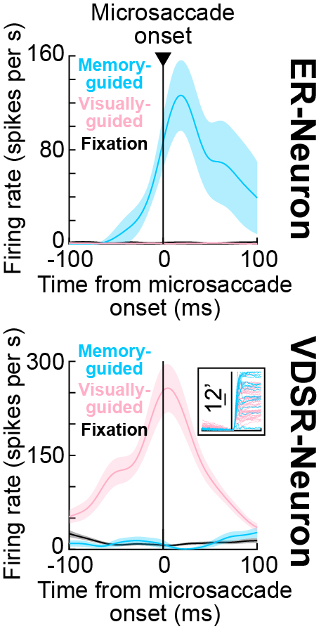 Specifically, one could find visual-only neurons, similar to our foveal SC neurons in this study, and one could also find microsaccade-movement-related discharge regardless of visual stimulation, like in this study. Most interestingly, we also found neurons showing either a movement-related discharge only in the presence of a visual stimulus (so-called visually-dependent microsaccade-related motor neurons); or, at the other end of the spectrum, neurons showing a movement-related discharge exclusively for memory-guided microsaccades. This functional diversity in the foveal region of the SC is amazing, and can lead to interesting questions about the role of such diversity in guiding behavior. The pre-print version of our paper can be read here.
