
We have recently performed extensive physiological studies of the foveal visual representation of the rhesus macaque superior colliculus (SC). As part of these studies, we have performed dense mappings of the retinotopic topography that is known to exist in this structure. Unlike in classic work, in which the mapping data were very sparse, we fully mapped the entire extent of the SC surface using very dense sampling. The result is that we now have a much more complete representation of SC topography, with much larger foveal magnification than that predicted by classic models. This has strong implications on views of the SC in orienting behavior. For example, it is classically assumed that the SC is a gaze-shifting structure (therefore concerned with only extra-foveal locations and receiving primarily magnocellular visual input). Our results recast this view.
To describe the complete SC topography, we fit a new model with the following parameters:
Bx = 1.1
By = 1.8
A = 0.9
These parameters are input into the following two equations, which convert visual locations (polar R and theta) into anatomical locations (X and Y in mm of neural tissue):
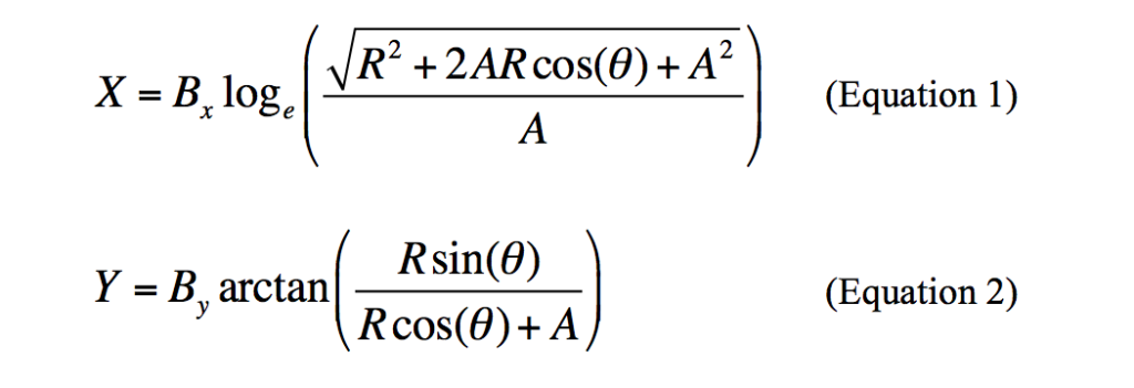
The result of the above two equations and our new fits (if you look down towards the SC from the top, as if recording in an experiment) is a map of visual space on the surface of the SC like the following:
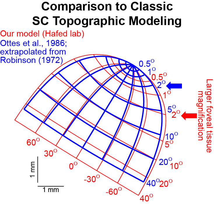
The red arrow in the figure above shows the 2 deg eccentricity line in the SC (towards the rostral end of the structure; i.e. the top end of the figure above). As can be seen, the foveal retinotopic region is very strongly magnified in the SC, at the expense of peripheral locations. To appreciate such magnification, one need only compare this new model to the original model, which was extrapolated from very sparse and peripheral measurements. Such comparison is provided in the figure below (red is the new model, and blue is the classic model), in which we aligned both models at the origin of the map (top-right corner of each map) to facilitate visual comparison. As can be seen, extrapolation of the sparse peripheral measurements in the classic model results in severe under-representation of the true amount of foveal magnification that is present in the SC. This can have implications on models that attempt to explain neurophysiological recordings in the SC:
We have also performed a 3-dimensional reconstruction of SC surface topography, such that we now have a 3-dimensional map. The details of this map are included in Data Table 1 below (also see the movies in the links below). The whole paper can be read online here:
- Chen, C.-Y., Hoffmann, K.-P., Distler, C., & Hafed, Z. M. (2019). The foveal visual representation of the primate superior colliculus. Current Biology, https://doi.org/10.1016/j.cub.2019.05.040. [PDF+SUPP INFO][SUPPLEMENTARY MOVIE 1][SUPPLEMENTARY MOVIE 2][DATA TABLE 1].