
Our lab has a new paper published in the Journal of Neurophysiology!
The superior colliculus (SC) of human and non-human primates receives a large part of its visual inputs via direct anatomical projections originating in the primary visual cortex (V1). Consistent with this, the visual responses of non-human primate SC neurons have long been known to appear, at least qualitatively, similar to those of V1 neurons. In our new work, we showed that such qualitative similarity between SC and V1 visual responses masks behind it a large and fundamental difference between the two brain areas when it comes to active vision.
Our manuscript addressed a known, yet perplexing, reality of brain organization: its massive “sensory parallelism”. For any given sensory input, information about this input is relayed relatively quickly, and essentially simultaneously, to several disparate parts of the brain. Such parallelism has been extensively discussed in the past from the perspective of sensory scene interpretation, with concepts like feature binding and dorsal-versus-ventral cortical processing streams receiving much research attention. However, sensory parallelism also touches intrinsically motor brain circuits. The question of why, say, an oculomotor control structure like the primate SC should also receive early sensory information has received less research effort to date, but this question is as important and fascinating as the questions of feature binding and differential cortical processing streams. This question is rendered even more urgent when past work has shown that sensory responses in both sensory (e.g. V1) as well as motor (e.g. SC) brain areas may still individually correlate to behavioral motor variability.
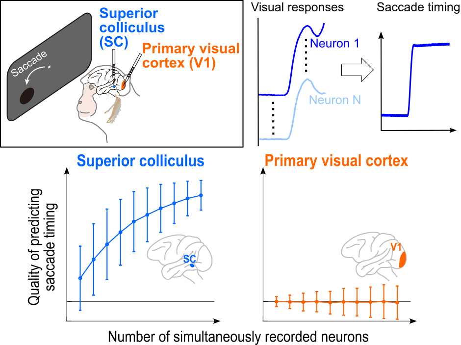
Our study investigated the contributions of visual response variability in either the SC or V1 to trial-by-trial variability in the timing of foveating eye movements. In our recent observations, made at the single-neuron level, we found that SC visual response variability was much more associated with behavioral variability than V1 visual response variability. For example, on trials with a weak SC visual response, saccadic reaction times were also later.
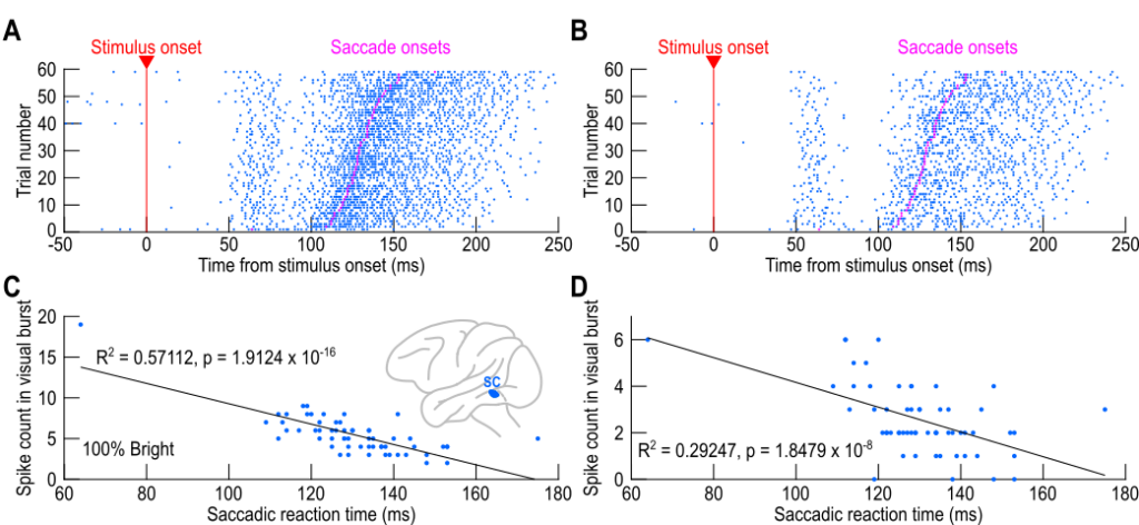
Our current study extended this line of work by investigating, for the first time, how well simultaneously recorded neurons in either the SC or V1 can predict trial-by-trial saccadic reaction time variability (using the same behavioral tasks, stimuli, and animals for both brain areas). We found that correlations between visual responses in V1 and trial-by-trial variability in foveation timing were virtually non-existent, even when considering populations of simultaneously recorded neurons. Thus, even at the population level, the differences between the SC and V1 that we previously observed persisted. Remarkably, the lack of relationship between V1 visual responses and saccadic eye movements was much stronger than we observed at the single-neuron level.

Our work in this new paper is related to a larger body of evidence from our lab, exploring fundamental differences between the SC and V1 when it comes to visual responses: like here, here, and here.