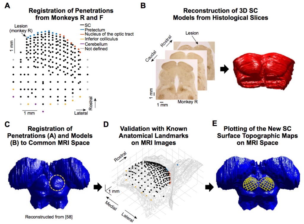
We have a new paper in press at Current Biology. In this paper, we did extensive neurophysiological and structural investigation of the foveal visual representation of the primate superior colliculus (SC).
The SC has historically been viewed, in primates, as primarily a gaze shifting structure. It was thus generally assumed that foveal visual scene analysis is the purview of retino-geniculate pathways, whereas gaze shifting away from the fovea is the function of the SC. However, we also make microscopic gaze shifts, which require visual guidance. Thus, even within foveal visual space, the SC must be involved. We therefore characterized the foveal visual representation of the SC.
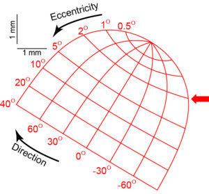
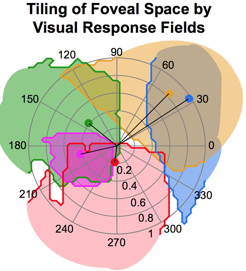
We found remarkable orderly organization in the SC’s representation of foveal visual input, and our topography measurements revealed that the SC contains much larger foveal magnification than previously anticipated. In fact, we found that the SC is as foveal a visual structure as primary visual cortex (V1)! This means that a large fraction of the amount of neurons in the SC is dedicated to only 1-2% of our entire visual field.
We also found that fixational eye movements, like microsaccades, are associated with altered foveal visual neuronal responses. These observations are a direct neural substrate for our human perceptual results in 2013 (Hafed, Neuron, 2013), in which we saw perceptual alteration around the time of microsaccades.
Finally, we found that slow ocular drifts cause major activations in SC visual activity.
The work was the result of a great collaboration with Klaus-Peter Hoffmann and Claudia Distler, from Bochum University.
An earlier BioRxiv version of the paper can be read here. The final published version can be read here.
Also, our new SC topography parameters can be reviewed here.

