Motor-related activity for microsaccades

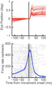
We recently identified neurons in the superior colliculus (SC) that exhibit motor-related activity for microsaccades (Hafed et al., 2009). The movie below demonstrates the activity of one such neuron, which exhibited preference for small upward eye movements. In the movie, the green graph shows a real-time representation of vertical eye position, and the white graph in the same panel shows horizontal eye position. As can be seen whenever the green graph deflected upward (upward microsaccade), the neuron burst (heard in the audio). In the panel under eye positions, we show a representation of radial eye velocity. Spikes in this velocity reflect microsaccadic eye movements. Finally, the panel below eye velocity shows a representation of firing rate of the neuron. The firing rate built up and burst for small, upward microsaccades, as shown in the figure on the left.
Visual activity in superior colliculus

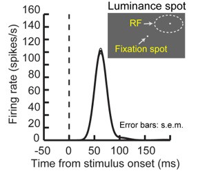
The following two movies show the activity of two SC neurons responding to peripheral visual stimuli. The neurons are generally silent and exhibit a short-lived visual response shortly after the presentation of stimuli inside their response fields, as demonstrated schematically on the right.
