Our lab combines neurophysiological and behavioral experiments with the techniques of real-time retinal-image stabilization and control.
Because the eyes continuously move, the brain engages in a perpetual visual-motor loop, in which eye movements cause changes in the location and motion of stimuli on the retina, and such changes drive subsequent eye movements, and so on. This creates an uncertainty about the state of retinal stimulation in a variety of experiments. For example, when we present a stationary stimulus on a display (like a spot or sine wave grating), even when subjects fixate, the eyes will move subtly, and this means that the stimulus becomes variable and random on the retina. However, if we use real-time control of the stimulus position that we present on the display, such that the position is dependent on the instantaneous state of eye movements, then this renders the retinal stimulus experimentally-controllable and less uncertain. This is a form of “perturbation experiment”, in which we perturb the normal operation of the visual system, by essentially playing tricks on it, in order to better understand how it operates. Combining this perturbation approach with neurophysiology is a particularly powerful approach that we try to exploit.
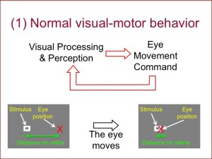
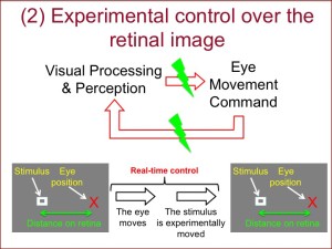
The principle for this technique can be understood from the two images below. In the first image (1), when the eye moves relative to a stationary stimulus, the distance between the fovea and the stimulus is altered on the retina. That is, the image of the stimulus changes location on the retina. However, in the second image (2), we perturb this expected effect of eye movement. For example, we move the stimulus on the display exactly with the position of the eye, such that on the retina, the stimulus stays at the same location.


As can be imagined, if we do indeed keep the stimulus at the same location on the retina, then there will always be a distance between the stimulus and the fovea. This distance is a powerful cue for the oculomotor system to generate an eye movement that attempts to bring the stimulus onto the fovea for closer inspection. As a result, a saccade will be triggered, but the stimulus will again be moved in real-time with the eye to keep the same retinal location as before. This will appear as if the stimulus is continuously attempting to escape the eye; the net result is a series of unstoppable saccades.
This phenomenon is shown in the video below. In this video, we show the eye position of a subject during this manipulation. As can be seen, the eye makes a series of rapid saccades, sometimes called “stair-case saccades”, because the eye position trace shows a series of “steps” in eye position. These stair-case saccades are a clear hallmark of the effect of the experimental manipulation that we applied. Of course, in real experiments, we apply this technique in much more subtle ways, such that the subject is not frustrated by the inability to foveate the stimulus. The video below is just an example demonstration of how powerful this technique can be in altering visual and eye movement behavior.
Retinal-image stabilization demo
Example studies employing this technique from our lab include (Chen and Hafed, 2013) and (Tian et al., 2016).
