We have a new article in press at the Journal of Neurophysiology. In this study, we explored the phenom enon of “saccadic suppression”, in which sensitivity to visual stimuli is reduced if these stimuli are presented around the time of saccadic eye movements. This phenomenon is normally thought to help us have a stable percept of our visual environment even though our eyes are in constant motion. However, despite being known for a very long time, the neural mechanisms for this phenomenon of saccadic suppression are not well understood.
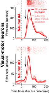
We investigated the neural mechanisms for saccadic suppression by recording activity from the superior colliculus (SC). This brain structure has two key properties that make it one of the possible brain sites for saccadic suppression: it has neurons that react to visual stimuli, and it also has neurons that generate an eye movement command. Thus, within the local SC circuit itself, a “corollary” of the movement command can be used to modulate the visual sensitivity of the visually-responsive neurons. In our study, we found that the visually-responsive neurons can indeed exhibit saccadic suppression. For example, Neurons A and B in the image on the right have lower visual sensitivity if a stimulus appears near a saccade (faint color) compared to when the same stimulus appears without an eye movement (saturated color).
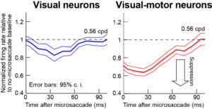
I nterestingly, not all visually-responsive SC neurons are created equal when it comes to saccadic suppression. Only a specific sub-type, called “visual-motor neurons” showed the strongest suppression, which was also time locked to the movement occurrence (see red image on the left). Moreover, the suppression depended on the stimulus spatial frequency characteristics, much like in perceptual tests of saccadic suppression in humans. Thus, it is only the visual-motor neurons that correlate with perceptual performance; other neurons (like the so-called SC “visual neurons”) have much milder saccadic suppression (see the blue image on the left). These results are particularly surprising and intriguing because previous studies have suggested that it would be the “visual neurons” that may indeed be the site for perceptually-related saccadic suppression in the SC, not the “visual-motor” neurons as we have found. Our results thus pave the way for further neurophysiological studies investigating this intriguing phenomenon associated with active perception.
