
 Whenever we maintain steady gaze at an object of interest, our eyes never remain perfectly stationary. Instead, they undergo several types of movements including ones designated as `microsaccades`. Microsaccades are extremely small versions of the saccadic eye movements that we employ in everyday life to scan our visual environment. These small movements were discovered several decades ago, but the brain mechanisms for generating them remained largely unknown. In a 2009 article in Science magazine, we uncovered evidence that the superior colliculus (SC), a brain structure important for specifying where we look, plays a crucial role in microsaccade generation. We have since followed up on these results with further investigations on the roles of these eye movements in visually-guided behavior.
Behavioral Characteristics of Microsaccades
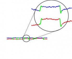
The following movies demonstrate some of the characteristics of microsaccades. In each movie, a representation of a subject`s eye position is shown in blue during a task in which the subject is instructed to maintain gaze at the center of the display. Notice how the eye moves continuously. Every so often, the subject makes a microsaccade, where the eye appears to abruptly change its position (indicated in the movies by a change in color from blue to red). As can be seen from the movies, these microsaccades happen frequently and have different directions and sizes. The gray circle in each movie is an approximate extent of the portion of our visual field in which we have relatively high acuity, and it provides a general idea of how small these microsaccades are. Also, note that the movies run in slow-motion so that these flicks in eye position can be seen more easily.
Neuronal activity in the superior colliculus associated with microsaccades
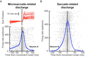 We found that individual neurons in the rostral portion of the superior colliculus increase their activity before and during microsaccades of a specific set of sizes and directions. We also found that different neurons have different preferences for these microsaccade metrics. For example, one neuron can preferentially represent movements to the right, and not be as active for leftward or upward movements, whereas another neuron can preferentially increase its activity for oblique (diagonal) movements but not for pure horizontal or vertical ones.
To illustrate this, the following movies show the same example trials above, but now with an additional representation of the activity of a single superior colliculus neuron recorded during the trials. Specifically, in each movie, every audible `click` indicates when the neuron emitted an action potential (more action potentials mean more neuronal activity). In addition, the cyan bar on the right has a height that is proportional to the instantaneous activity of the neuron. As can be seen (and heard), this particular neuron systematically increased its activity before and during microsaccades with a rightward component, and it reduced its activity (or was unmodulated) before and during other microsaccades. Thus, even from just a single trial, the neuron`s preference for some microsaccades and not others is evident.