
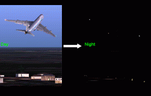
Physiological studies of oculomotor control have historically relied on individual spots of light as the visual targets. However, in everyday life, we often orient towards extended objects that may or may not be moving. For example, the above scene on the left involves a large object that we can smoothly track by directing our gaze to its center. At night (above right), the same scene has a sparser visual representation of the object (only the strobe lights on the wing tips may be visible), but we are still able to track the object`s invisible center. This latter scenario is tractable for study in the laboratory, which allowed us to investigate some of the neural mechanisms associated with it. We have described these mechanisms using single-neuron recording from and reversible inactivation of the superior colliculus (SC), a structure crucial for oculomotor control. The following are sample demonstrations of the basic results we obtained.
Sample Behavior
The following movies (running at half speed) show sample stimulus configurations, along with a representative subject`s eye position indicated by a green crosshair. During `extrafoveal tracking`, the subject successfully inferred the invisible center of a stimulus consisting of two peripheral features and tracked it for several seconds.
Behavior of Individual SC Neurons
We recorded from superior colliculus (SC) neurons during the above behaviors. The SC contains a topographic representation of visual space, which allowed us to record either from peripheral neurons (representing one of the visual features of the stimulus) or central ones (representing the foveal location of the invisible center that was tracked). We found that the central neurons were the most active during extrafoveal tracking, despite the lack of a visual stimulus. These neurons were modulated based on the location of the goal that the subject was trying to orient towards. During fixation, when the invisible center could be ignored, the neurons were modulated to a lesser extent. The following movies illustrate some of these findings.
- Sample SC neuron from the central portion of the SC during fixation (the graph on the bottom is an indication of the neuron`s activity) (Quicktime format, running at half speed)
- The same neuron during extrafoveal tracking (the gray curve in the bottom is the activity during fixation shown in the above movie) (Quicktime format, running at half speed)
Effects of Reversible Inactivation of a Subset of SC Neurons
The following movie shows a representation of extrafoveal tracking performance with and without SC inactivation. The white crosshair shows the subject`s average eye position from a baseline data set. The yellow crosshair shows this position after locations in the SC encoding the lower left quadrant (in retinotopic coordinates) were reversibly inactivated. The subject tracked as in the baseline condition, but with a constant offset (relative to baseline) to the upper right quadrant, indicating a biased estimate of goal location.
- Effects of reversible inactivation on extrafoveal tracking (Quicktime format, running at half speed)