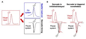
 Our lab has a new paper published in the Journal of Neurophysiology, in which we investigated the potential mechanisms for controlling microsaccades. These tiny eye movements occur frequently during gaze fixation, and we have previously shown that the superior colliculus (SC), a brainstem oculomotor structure, is involved in their generation. In the latest experiments, we explored what happens when visual stimuli are presented simultaneously with the generation of microsaccades. Because the SC has a topographic map of spatial locations, these scenarios result in the presence of two simultaneous “hills” of neural activity at microsaccade generation: a hill representing the movement goal location, and a second hill representing the visual stimulus location. Depending on the direction of the microsaccade and the location of the visual “hill” of neural activation, we hypothesized that the two hills might compete to either boost microsaccade size or reduce it.
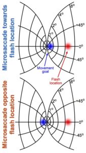
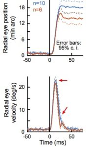 We indeed found that a microsaccade congruent with stimulus location (i.e. “towards” the stimulus) was bigger than a microsaccade “opposite” the stimulus. Interestingly, this happened even though the peak speed of eye rotation during both microsaccades was identical. This result is intriguing because it represents an example of a violation of a well-known “rule” in the saccadic system, in which peak velocity increases with movement amplitude. Based on this observation, we developed a simple model of oculomotor control that attempts to explain our findings.
Our work allows testable hypotheses about eye movement generation in the brain. More broadly, since eye movements are a window on the mind, and can be important clinical indicators of neurological disorders, understanding eye movement control can be tremendously useful for both cognitive neuroscience and clinical applications.